WHOOPS!
404
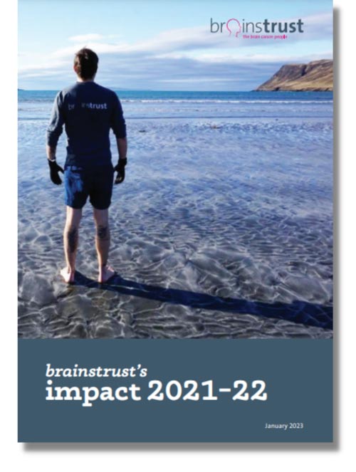 Our vision is for everyone with a brain tumour to feel less afraid, less alone and more in control.
Last year your support helped:
2,339 people contacted our team of support specialists for help
825 new people accessed our support
404 people receive a Brain Box
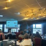
We hosted 134 support events
110,928 people access our online information and support
3200 people in our online community to help each other to feel less alone.